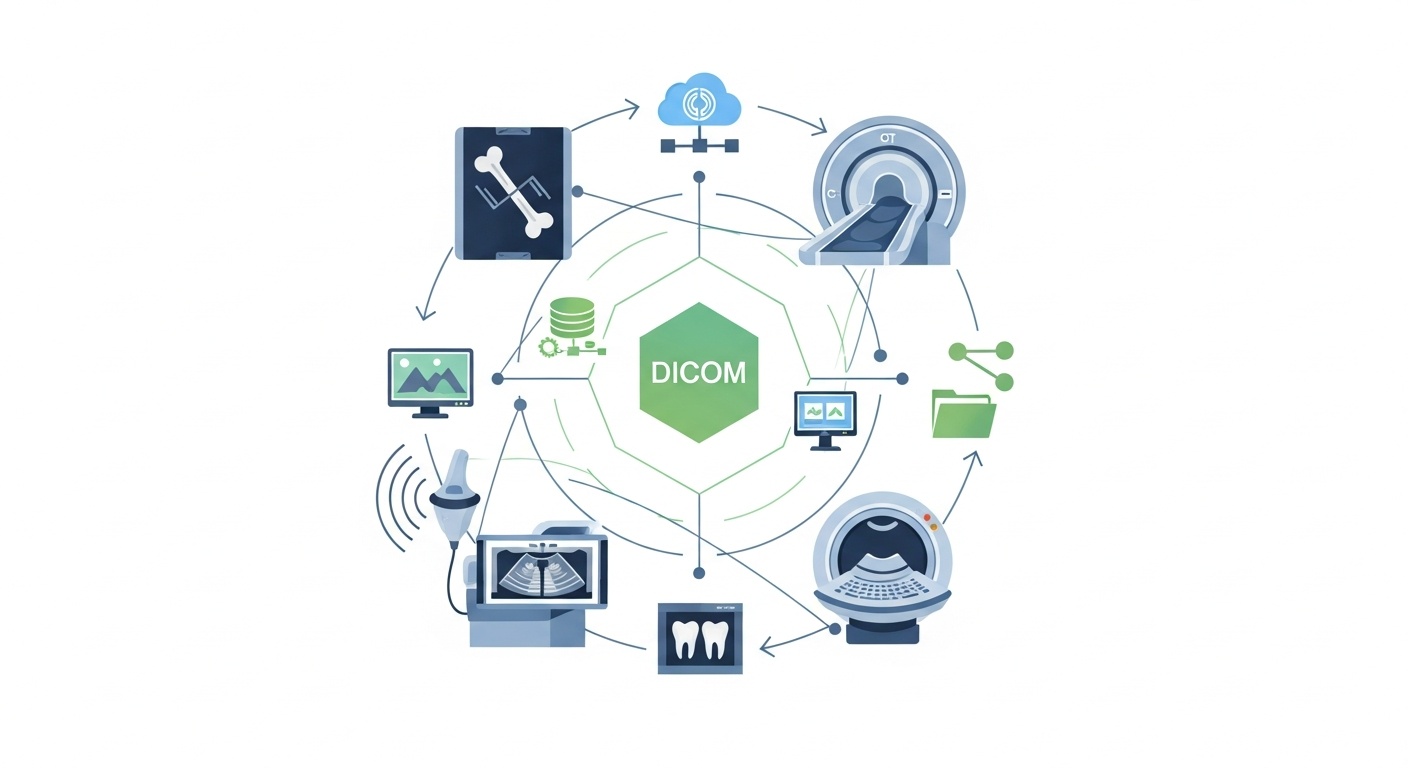
Today, veterinary imaging is a standard practice in veterinary medicine. Clinics and referral hospitals rely on our X-rays, CTs, MRIs, Ultrasounds, dental imaging, and other modalities to diagnose injuries and disease and to help determine the best treatment. As these imaging exams get more sophisticated, veterinary teams must be able to access, share, store, and manage these exams in a consistent manner. This is where the role of DICOM is important.
In veterinary medicine, DICOM is the standard format for the storage, presentation, transmission, and sharing of medical imaging studies of animals, including X-rays, computed tomography (CT) scans, magnetic resonance imaging (MRI) scans, ultrasound, and dental images.
It enables veterinary clinics to integrate imaging devices, image viewers, PACS systems, and specialist referral processes. DICOM is not a diagnostic tool used on its own, but it provides a consistent method for accessing and reviewing imaging studies by veterinarians and veterinary radiologists.
DICOM is the acronym for Digital Imaging and Communications in Medicine. It has many applications in human medicine and some in veterinary radiology and imaging. A DICOM file may contain the image, patient information, study information, modality type, scan date, and other information that is useful for organizing the study.
This information can be used in veterinary medicine, such as the animal's name, species, breed, owner details, clinic information, and imaging study data. This facilitates easier identification, retrieval, comparison, and sharing of animal imaging research studies.
The DICOM standard for medical imaging is helpful because it enables different medical imaging devices and software systems to operate with a more consistent format for medical images.
Animal imaging is not as consistent as human imaging. The positioning, imaging techniques, and interpretation methods may be different for a cat, dog, horse, rabbit, or exotic bird. Veterinary teams also have to deal with images from various devices, and in some cases, transfer images to outside experts.
One way DICOM helps in this process is by providing a structured image format that can be used on compatible systems within the clinic. Veterinary practices can store and manage studies efficiently, rather than just on CDs or a single workstation, or through USB drives.
DICOM can assist veterinary teams to:
• Group Imaging Studies By Visit & Patient.
• Look At Previous Research To Compare With.
• Examine Pictures Of Various Modalities
• Conduct Studies In Collaboration With Specialists Or Referral Hospitals.
• Minimize Reliance On Physical Media
• Ensure The Long-term Preservation Of Images In The Archive.
The final diagnosis will also rely on the veterinarian/veterinary radiologist, the animal's history, clinical signs, and the examination.
Depending on the imaging system and software configuration, many systems will be able to generate or export DICOM images.
| Imaging modality | Common veterinary use | How DICOM helps |
| X-ray / Digital Radiography | Bone injuries, chest imaging, abdominal review | Stores images in a consistent format for viewing and comparison |
| CT | Trauma cases, complex bone conditions, tumor evaluation, and surgical planning | Supports detailed cross-sectional image review |
| MRI | Brain, spine, neurological, and soft tissue evaluation | Allows organized review of advanced imaging studies |
| Ultrasound | Abdominal, cardiac, reproductive, and soft tissue imaging | Stores selected images or cine loops with study details |
| Dental imaging | Tooth roots, jaw structure, and oral disease evaluation | Keeps dental images connected to the patient record |
This doesn't imply that all devices have the same workflow. The exact process will vary depending on the available imaging equipment, the clinic setup, and the software being used.
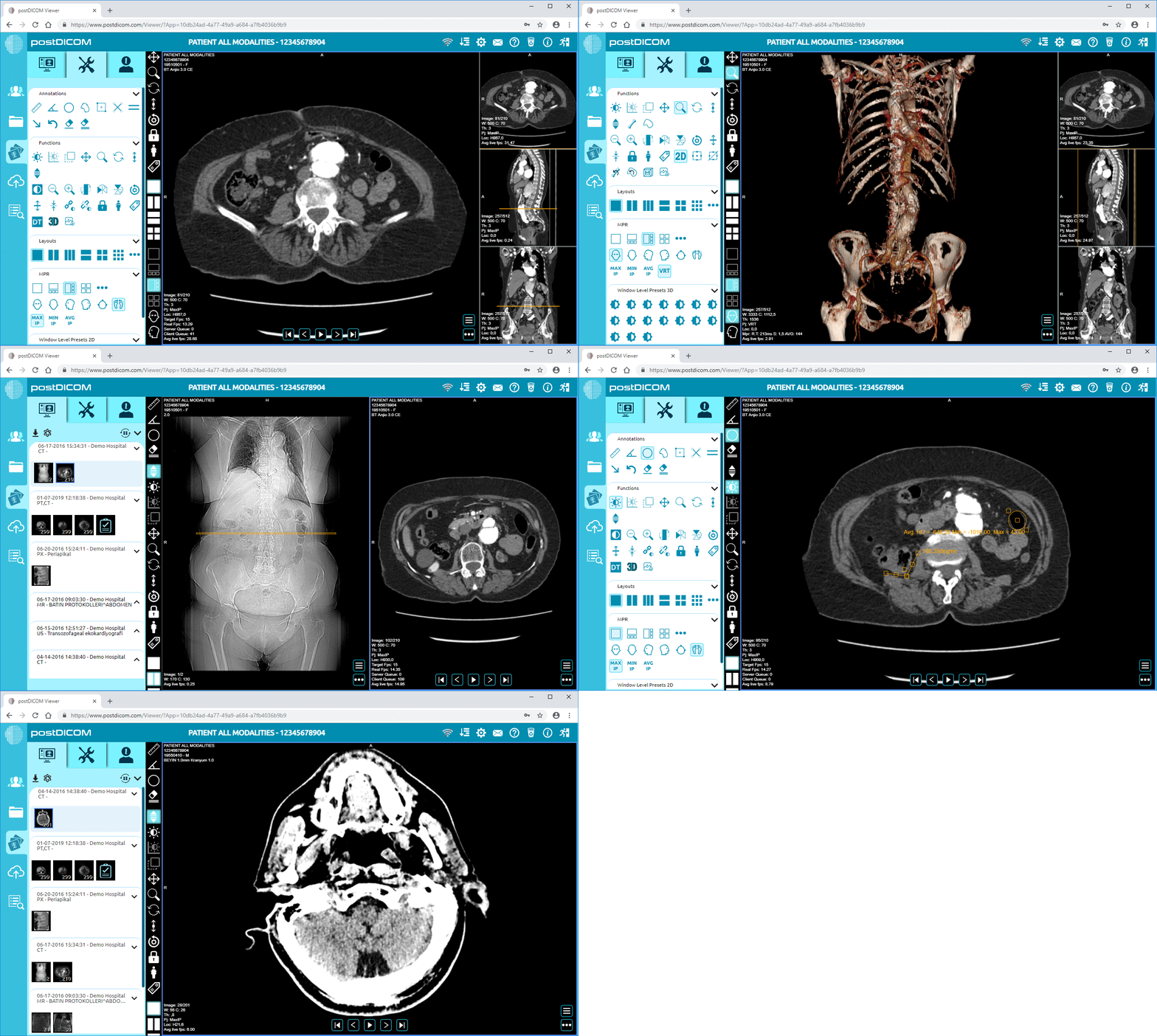
 - Created by PostDICOM.jpg)
There are connections between DICOM, PACS and the DICOM viewers, but they all have different roles.
| Term | What it means | Veterinary example |
| DICOM | The standard format for imaging studies and related data | A dog’s CT scan saved as a DICOM study |
| PACS | A system for storing and managing imaging studies | A clinic stores animal scans in a digital archive |
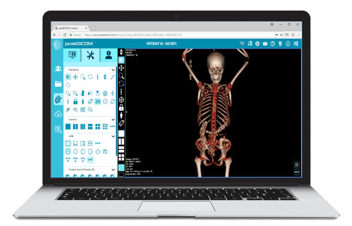
| DICOM viewer | Software used to open and review DICOM images | A veterinarian reviews an MRI study in a browser-based viewer |
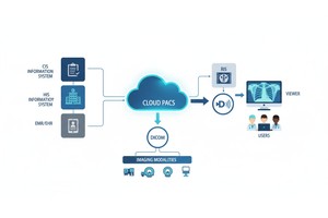
A common veterinary imaging workflow might have an imaging device, PACS, and then a viewer open for the veterinarian to view the study. With a modern diagnostic DICOM viewer, the veterinary team can access and view all of these images without having to rely on just one workstation.
Many veterinary practices have partners and specialists with whom they collaborate. Images may be sent from a general clinic to a veterinary radiologist, surgeon, neurologist, or referral hospital for review. This is frequently found in orthopedic, neurological, oncology, dental, and emergency surgeries.
DICOM makes this possible by enabling sharing of the imaging study in a format that is readable by compatible systems. Clinics can employ a digital sharing workflow system rather than sending large files through an insecure channel or mailing discs.
Secure medical image sharing can provide secure access to medical images from clinics without altering the image files. This also contributes to veterinary teleradiology, in which veterinary imaging studies are remotely examined by a qualified veterinary imaging specialist.
DICOM can be used in various veterinary practices.
Digital X-ray can be used on dogs and cats at a small-animal clinic. Previous images can also be recalled when a patient returns for follow-up care and can be used to make a clinical comparison with the newer studies.
Complex cases may require referral to a facility to perform a CT or MRI. These studies typically consist of numerous image slices, and with this, a well-organized file system and sophisticated viewing software can streamline the review process.
Imaging can be used in an equine clinic to evaluate lameness, trauma, and dental issues, and for follow-up. With the organization of studies it will be easier for the care team to see the imaging history over time.
A multi-location veterinary group may require access to imaging studies from various locations. Authorized users may find it easier to view studies using cloud storage and browser-based viewing.
A typical workflow for a veterinary DICOM system would be:
1. X-ray, Ct Scan, Mri, Ultrasound, Dental Imaging Or Other Method Are Used To Scan The Animal.
2. A Dicom Study Is Created Or Exported By The Imaging Device.
3. The Study Is Submitted To A Pacs Or Cloud Storage System.
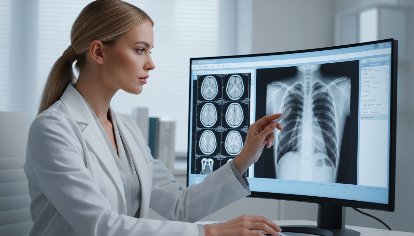
4. The Veterinarian Opens The Study In A Dicom Viewer.
5. The Clinic Communicates The Study With The Specialist As Required.
6. The Study Continues To Be Available For Future Review Or Comparison.
In such practices, where browser based access, secure storage and ease of image sharing is required, a veterinary cloud PACS can be of assistance. It is particularly useful for clinics which have to do with referral partners, outside radiologists or a number of imaging devices.
There are some practical issues with the workflow of veterinary imaging. Animal patients may move during the scan, positioning may be challenging and anatomy varies from species to species. Clinics can also employ various imaging equipment made by various manufacturers.
Common challenges include:
• Controlling Images Over Several Devices
• Transferring Large Imaging Studies To Specialists
• The Studies Were Organised By Animal, Owner And Visit.
• The Need For Older Research For Comparison
• Minimizing Dependence On Cds Or Local Computers.
• Supporting Access To Multiple Clinic Sites
Some of these can be mitigated with the help of DICOM viewers, PACS systems, and image sharing software. The ideal setup, however, will differ depending on the size of the clinic, the number of images being captured, the efficiency of the workflow and the specialty referral requirements.
DICOM is an acronym for Digital Imaging and Communications in Medicine. In veterinary medicine, it is the standard format for the storage, display, transfer, and management of imaging studies of animals.
Veterinary practices that utilize digital imaging equipment like X-ray, CT, MRI, dental imaging or ultrasound might require a DICOM viewer. A DICOM viewer lets vets open and view imaging studies as needed.
No. DICOM is widely used in human healthcare, but it can also be used in veterinary imaging. The DICOM standard can handle imaging studies in animal hospitals, specialty clinics and veterinary radiologists.
DICOM facilitates the exchange of imaging studies between clinic systems that are compatible with each other. This can assist with specialism review, second opinions, referrals, consultations and remote radiology workflows.
Yes. A cloud-based PACS can be set up by many veterinary practices to store imaging studies. The appropriate configuration will be determined by the imaging equipment, workflow, privacy and sharing requirements at the clinic.
DICOM assists the veterinary team to perform animal imaging studies more organized and consistently. Stores, views, shares, refers and compares images in various modalities (X-ray, CT, MRI, ultrasound and dental imaging).
DICOM is more than just a technical file format for veterinary clinics. It is included in a practical imaging workflow that assists veterinarians in accessing studies, collaborating with specialists, and more effectively managing animal patient records.
|
Cloud PACS and Online DICOM ViewerUpload DICOM images and clinical documents to PostDICOM servers. Store, view, collaborate, and share your medical imaging files. |