Imagine a world where your medical team can explore the human body in vivid detail from every conceivable angle with just a few clicks. Picture being able to transform flat, two-dimensional scans into dynamic, three-dimensional models that bring clarity to the most complex cases.
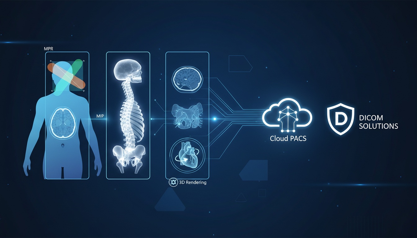
This isn't the future—it's the present, thanks to advanced image processing tools like Multi-Planar Reconstruction (MPR), Maximum Intensity Projection (MIP), and 3D rendering.
These tools are game-changers for medical facilities, research institute owners, and managers. They streamline clinical workflows, improve diagnostic accuracy, and enhance patient care. But the real magic happens when these tools are seamlessly integrated into a robust platform like PostDICOM’s Cloud PACS and DICOM solutions.
In this blog post, we’ll explore advanced image processing, including how MPR, MIP, and 3D rendering work, why they’re essential, and how PostDICOM’s cutting-edge platform brings these technologies to life.
Join us as we uncover the transformative potential of these tools and how they can revolutionize your clinical workflow.
Multi-planar reconstruction (MPR) is an advanced imaging technique that allows radiologists and medical professionals to view cross-sectional images of a particular body part in multiple planes.
Typically derived from CT or MRI scans, MPR enables the visualization of structures from various perspectives: axial (horizontal), sagittal (side), and coronal (frontal).
Detailed Anatomical View: MPR is crucial for viewing complex anatomical structures, such as the brain, spine, and joints. It provides a comprehensive understanding by displaying different planes, aiding in diagnosing conditions like fractures, tumors, and congenital abnormalities.
Surgical Planning: Surgeons use MPR to plan procedures more effectively. Examining the anatomy from multiple angles allows them to anticipate challenges and strategize their approach, improving surgical outcomes.
Oncology: MPR is particularly valuable for assessing tumor size, shape, and extent. It provides a clear, multi-dimensional view of the affected area, helping track tumor progression and plan radiation therapy.
Maximum Intensity Projection (MIP) is an image processing technique used to visualize high-intensity structures within volumetric data. MIP projects the voxel with the highest intensity along a specific viewing direction onto a 2D image, making it ideal for highlighting structures like blood vessels, airways, and other high-contrast regions.
Vascular Imaging: MIP is extensively used in angiography to visualize blood vessels. It provides clear and detailed images of the vascular network, helping to detect vascular diseases such as aneurysms, stenosis, and blockages.
Pulmonary Imaging: In thoracic imaging, MIP enhances the visibility of pulmonary nodules and other lung pathologies, aiding in the early detection and treatment of respiratory conditions.
Neurology: MIP is used in brain imaging to highlight vascular structures and detect abnormalities like arteriovenous malformations (AVMs) and intracranial hemorrhages.
3D rendering is an advanced technique that converts two-dimensional cross-sectional images into three-dimensional models. This process involves sophisticated algorithms that reconstruct the 3D structure of an object from multiple 2D slices, providing a detailed and realistic view of the anatomy.
Surgical Planning: 3D rendering is invaluable in surgical planning. Surgeons can manipulate the 3D model to examine the anatomy from all angles, identify potential challenges, and plan their approach more precisely.
Diagnostic Evaluation: 3D models provide a more intuitive understanding of complex anatomical relationships, enhancing the accuracy of diagnoses. They are instrumental in orthopedics, cardiology, and oncology.
Patient Education: 3D-rendered images are powerful tools for patient education. They help patients visualize and understand their conditions, treatment plans, and surgical procedures, improving communication and compliance.
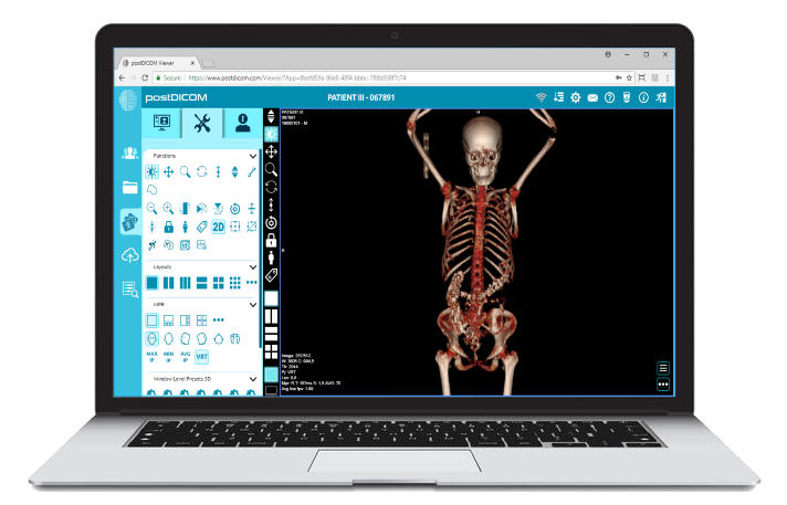
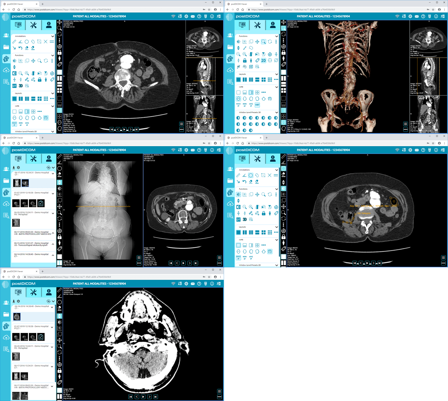
PostDICOM’s platform excels in integrating advanced image processing tools like MPR, MIP, and 3D rendering, providing a comprehensive solution for medical imaging. PostDICOM enhances diagnostic accuracy, streamlines clinical workflows, and improves patient care by offering intuitive interfaces, customizable options, and seamless workflow integration.
PostDICOM’s platform offers a user-friendly interface that makes Multi-Planar Reconstruction (MPR) easily accessible and highly functional:
Intuitive Navigation: Users can effortlessly navigate through different planes (axial, sagittal, and coronal) with simple controls, ensuring a smooth and efficient workflow.
Interactive Display: The interactive display allows users to adjust the plane of reconstruction dynamically, allowing them to focus on specific areas of interest for detailed examination.
PostDICOM provides extensive customization options for MPR:
Adjustable Parameters: Users can modify parameters such as slice thickness and spacing, contrast, and brightness to tailor the images to their diagnostic needs.
Annotation Tools: Built-in annotation tools enable healthcare professionals to mark areas of interest, add notes, and share insights with colleagues, enhancing collaborative diagnosis and treatment planning.
The integration of MPR in PostDICOM’s platform offers several clinical benefits:
Enhanced Diagnostic Accuracy: MPR improves accuracy by allowing clinicians to view anatomical structures from multiple perspectives.
Streamlined Workflow: The seamless integration of MPR tools into the platform reduces the time spent switching between different software, facilitating the diagnostic process and improving efficiency.
PostDICOM’s platform includes powerful tools for Maximum Intensity Projection (MIP):
High-Quality Visualization: MIP images are rendered with high fidelity, ensuring that the highest-intensity structures, such as blood vessels and pulmonary nodules, are visible.
Customizable Views: Users can adjust the viewing angle and projection parameters to optimize the visualization of specific structures, enhancing the clarity and diagnostic value of the images.
MIP is seamlessly integrated into PostDICOM’s clinical workflows:
Automatic Generation: MIP images can be automatically generated from volumetric data, saving time and effort for healthcare professionals.
Easy Sharing: The platform allows for sharing MIP images with other specialists, facilitating collaborative diagnosis and treatment planning.
Real-world examples highlight the impact of MIP integration in PostDICOM:
Vascular Imaging: A case study in a cardiology department showed that using PostDICOM’s MIP tools improved the detection of coronary artery blockages, leading to more timely and effective interventions.
Pulmonary Imaging: In a pulmonology clinic, MIP images generated by PostDICOM’s platform enhanced the visibility of small nodules, aiding in the early detection of lung cancer.
PostDICOM supports various advanced 3D rendering techniques:
Volume Rendering: This technique provides a comprehensive view of the internal structure of the anatomy and is helpful for complex diagnostic evaluations.
Surface Rendering: Surface rendering highlights the outer surfaces of structures, which is particularly useful in surgical planning and orthopedics.
PostDICOM’s 3D rendering tools include interactive features that enhance image analysis:
Rotation and Zoom: Users can rotate and zoom into the 3D models to examine details from different angles, providing a thorough understanding of the anatomy.
Slicing and Cross-Sectioning: The platform allows users to slice through 3D models to view internal structures, facilitating detailed examination and diagnosis.
The clinical impact of 3D rendering with PostDICOM is significant:
Surgical Planning: Surgeons can use 3D models to plan procedures more precisely, improving surgical outcomes and reducing risks.
Patient Communication: 3D-rendered images are practical tools for explaining conditions and treatment plans to patients, enhancing their understanding and engagement.
PostDICOM’s platform delivers comprehensive image analysis by integrating advanced image processing tools into a unified, user-friendly, and secure system.
With seamless integration, cross-modality support, an intuitive design, customizable workflows, and robust security measures, PostDICOM enhances the efficiency and effectiveness of clinical workflows.
PostDICOM’s platform is designed to integrate seamlessly with a wide range of imaging modalities and medical devices, providing a unified solution for comprehensive image analysis:
Multi-Modality Support: The platform supports various imaging modalities, including CT, MRI, PET, ultrasound, and X-ray. This cross-modality compatibility ensures that all relevant imaging data can be accessed and analyzed within a single system.
Centralized Access: By consolidating imaging data from multiple sources into one centralized platform, PostDICOM eliminates the need for disparate systems, streamlining the workflow and enhancing efficiency.
With PostDICOM, users can seamlessly handle images from different modalities within the same analysis framework:
Integrated View: The platform allows users to view and compare images from different modalities, providing a holistic view of the patient’s condition. This capability is precious in complex cases where multi-modality imaging is required for accurate diagnosis and treatment planning.
Unified Analysis Tools: PostDICOM provides advanced analysis tools that can be applied across all supported modalities, ensuring consistent and comprehensive image evaluation.
PostDICOM places a strong emphasis on user experience, offering an intuitive interface that enhances usability across different devices:
Consistent Experience: The platform’s design ensures a consistent user experience, whether accessed from a desktop, tablet, or smartphone. This consistency reduces the learning curve and enables healthcare professionals to work efficiently across various devices.
User-Friendly Controls: The interface features user-friendly controls for navigating and manipulating images, making it easy for users to perform detailed analyses and annotations.
PostDICOM’s platform is highly customizable, allowing users to tailor workflows to meet specific clinical needs:
Flexible Settings: Users can customize viewing preferences, annotation tools, and analysis parameters to match their unique requirements. This flexibility ensures the platform can adapt to diverse clinical environments and practices.
Optimized Efficiency: By enabling users to create and save custom workflows, PostDICOM helps maximize efficiency, reducing the time spent on routine tasks and allowing healthcare professionals to focus on patient care.
PostDICOM prioritizes the security of patient data, implementing robust measures to protect sensitive information:
Encryption: All data stored and transmitted through PostDICOM’s platform is encrypted, ensuring it remains confidential and secure.
Access Controls: The platform employs stringent access control mechanisms, allowing administrators to define user permissions and ensure that only authorized personnel can access or modify medical images.
PostDICOM’s solutions are designed to provide seamless access across multiple devices, supporting flexible and dynamic workflows:
Anywhere, Anytime Access: Healthcare professionals can access medical images from any location and on any device, whether in the office, at home, or on the go. This flexibility is crucial for timely decision-making and continuous patient care.
Responsive Design: The platform’s responsive design ensures that images are displayed clearly and accurately on all devices, from desktops to tablets and smartphones. This allows users to perform detailed analyses regardless of their device.
 - Created by PostDICOM.jpg)
These case studies and real-world examples highlight the transformative impact of PostDICOM’s advanced image-processing tools and unified platform.
By enhancing diagnostic accuracy, optimizing surgical planning, and facilitating remote consultations, PostDICOM helps healthcare providers deliver high-quality care, improve clinical workflows, and achieve better patient outcomes.
Background
A major metropolitan hospital sought to improve diagnostic accuracy and streamline imaging workflows. However, fragmented systems and a lack of advanced image processing tools hindered the efficiency of its diagnostic processes.
Solution
The hospital implemented PostDICOM’s Cloud PACS and DICOM solutions, leveraging advanced tools like Multi-Planar Reconstruction (MPR), Maximum Intensity Projection (MIP), and 3D rendering for comprehensive image analysis.
Results
Improved Diagnostic Precision: PostDICOM’s advanced image processing tools allowed radiologists to examine complex anatomical structures from multiple angles, significantly enhancing diagnostic precision.
Streamlined Workflows: The unified platform allowed seamless integration of images from various modalities, reducing the time spent on image retrieval and analysis.
Enhanced Collaboration: Multi-device compatibility enabled specialists to collaborate in real-time, regardless of location, improving diagnosis accuracy and speed.
Testimonial
Dr. Sarah Thompson, Chief Radiologist, stated, “PostDICOM’s platform has revolutionized our diagnostic processes. The ability to view and manipulate images from different modalities in one place has greatly improved our efficiency and diagnostic accuracy.”
Background
An orthopedic clinic needed a solution to enhance its surgical planning capabilities. The clinic’s existing imaging systems were inadequate for detailed preoperative planning, particularly for complex orthopedic surgeries.
Solution
The clinic adopted PostDICOM’s Cloud PACS and advanced 3D rendering tools to improve its surgical planning processes.
Results
Detailed 3D Models: Surgeons were able to create detailed 3D models of patients’ anatomy, providing a comprehensive view that was essential for planning complex surgeries.
Improved Outcomes: 3D rendering for preoperative planning led to more precise surgical interventions, reducing the risk of complications and improving patient outcomes.
Patient Communication: The 3D models were also used to explain surgical procedures to patients, enhancing their understanding and engagement in treatment.
Testimonial
Dr. John Miller, an Orthopedic Surgeon, commented, “The 3D rendering capabilities provided by PostDICOM have been a game-changer for our surgical planning. We can plan our procedures more precisely and communicate more effectively with our patients.”
Background
A rural health network faces challenges in providing timely and comprehensive care due to geographical barriers and a shortage of specialists. The network aimed to leverage telemedicine to overcome these challenges but needed a robust solution for remote access to medical images.
Solution
The health network implemented PostDICOM’s Cloud PACS and multi-device compatible DICOM viewers to support its telemedicine initiatives.
Results
Remote Access: Physicians could access and review medical images remotely from their tablets and smartphones, ensuring continuous and comprehensive patient care.
Specialist Consultations: The network facilitated remote consultations with specialists who could review images and provide second opinions from their own devices, improving the quality of care for patients in remote areas.
Timely Interventions: Accessing updated images quickly allowed for timely interventions, reducing the risk of complications and improving patient outcomes.
Testimonial
Dr. Emily Harris, a General Practitioner, noted, “PostDICOM’s Cloud PACS has greatly enhanced our telemedicine services. Accessing and sharing images remotely has improved our patient care immensely, allowing us to provide specialist-level care even in rural communities.”
Advanced image processing tools like Multi-Planar Reconstruction (MPR), Maximum Intensity Projection (MIP), and 3D rendering are revolutionizing medical imaging.
These technologies provide healthcare professionals with unparalleled insights, allowing for more accurate diagnoses, better treatment planning, and enhanced patient care. Understanding the capabilities and applications of these tools is essential for any medical facility looking to stay at the forefront of medical innovation.
PostDICOM’s Cloud PACS and DICOM solutions seamlessly integrate MPR, MIP, and 3D rendering into a unified platform, making these powerful tools accessible and easy to use. With PostDICOM, medical professionals can leverage advanced image processing to improve clinical workflows, facilitate collaboration, and ensure they provide the best possible care to their patients.
By incorporating these advanced tools, PostDICOM enhances diagnostic accuracy, streamlines surgical planning, and supports comprehensive remote consultations. The real-world examples and case studies discussed demonstrate these technologies' significant impact on improving healthcare outcomes.
Explore PostDICOM’s advanced imaging solutions today to experience the transformative power of MPR, MIP, and 3D rendering and elevate your clinical workflows to new heights. Whether you are a medical facility, a research institute, or a healthcare manager, PostDICOM provides the tools you need to stay ahead in the ever-evolving world of medical imaging.


|
Cloud PACS and Online DICOM ViewerUpload DICOM images and clinical documents to PostDICOM servers. Store, view, collaborate, and share your medical imaging files. |